Pancreatitis 4s5l5x
This document was ed by and they confirmed that they have the permission to share it. If you are author or own the copyright of this book, please report to us by using this report form. Report 3b7i
Overview 3e4r5l
& View Pancreatitis as PDF for free.
More details w3441
- Words: 1,717
- Pages: 7
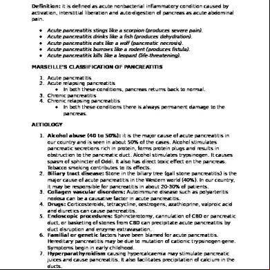
ACUTE PANCREATITIS Definition: it is defined as acute nonbacterial inflammatory condition caused by activation, interstitial liberation and auto-digestion of pancreas as acute abdominal pain.
Acute Acute Acute Acute Acute
pancreatitis pancreatitis pancreatitis pancreatitis pancreatitis
stings like a scorpion (produces severe pain). drinks like a fish (produces dehydration). eats like a wolf (pancreatic necrosis). burrows like a rodent (produces fistula). kills like a leopard (life-threatening).
MARSEILLE’S CLASSIFICATION OF PANCREATITIS 1. Acute pancreatitis 2. Acute relapsing pancreatitis In both these conditions, pancreas returns back to normal. 3. Chronic pancreatitis 4. Chronic relapsing pancreatitis In both these conditions there is always permanent damage to the pancreas. AETIOLOGY 1. Alcohol abuse (40 to 50%): it is the major cause of acute pancreatitis in our country and is seen in about 50% of the cases. Alcohol stimulates pancreatic secretions rich in protein, forms protein plugs and results in obstruction to the pancreatic duct. Alcohol stimulates trypsinogen. It causes spasm of sphincter of Oddi. It also has direct toxic effect on the pancreas. Tobacco smoking contributes to its effects. 2. Biliary tract disease: Stone in the biliary tree (gall stone pancreatitis) is the major cause of acute pancreatitis in the Western world (40%). In our country, it may be responsible for pancreatitis in about 20-30% of patients. 3. Collagen vascular disorders: Autoimmune disease such as polyarteritis nodosa can be a causative factor in acute pancreatitis. 4. Drugs: Corticosteroids, tetracycline, oestrogens, azathioprine, valproic acid and diuretics can cause pancreatitis. 5. Endoscopic procedures: Sphincterotomy, cannulation of CBD or pancreatic duct, or basketing of stones from CBD can precipitate acute pancreatitis by duct disruption and enzyme extravasation. 6. Familial or genetic factors have been blamed for acute pancreatitis. Hereditary pancreatitis may be due to mutation of cationic trypsinogen gene. Symptoms begin in early childhood. 7. Hyperparathyroidism causing hypercalcaemia may stimulate pancreatic juices and cause pancreatitis. It also facilitates precipitation of calcium in the ducts.
8. Hyperlipidaemia (>1000mg/dl of triglycerides) can also cause pancreatitis. Dietary control can cure ischaemia to the pancreas resulting in acute pancreatitis. 9. Hypothermia and hypotension can cause ischaemia to the pancreas resulting in acute pancreatitis. 10.Injury to the pancreas either postoperative or following penetrating injury can result in pancreatitis. 11.Infection: Virus such as Mumps and Coxsackie can cause pancreatic. Scorpion sting can alson cause pancreatitis abnormality. 12.Ductal: Around 20%. Some of them are due to pancreatic divisum seen during ER. The opeing of the minor papilla is inadequate for drainage of pancreatic juice. 13.Postoperative pancreatitis It follows operations on CBD-open or laparoscopic. More common after T-tube insertions (now it is less). Sphincteroplasty, ER, stone extractions Pancreatic biopsy Gastrectomy (distal/total) Cardiac surgery. Here risk factors are perioperative istration of calcium, postoperative hypotension, preoperative renal failure. 14.Idiopathic: It is seen in about 15% of cases. Even though, classified as idiopathic (no cause is found), they are found to have sludge/gall stones undetected by ultrasound examination. PATHOGENESIS Autodigestion is the final common pathway leading to pancreatitis; a few theories have been explained as possible factors for autogenesis. 1. Obstruction/secretion: Recurrent attacks of pancreatitis as in chronic cases can be due to multiple strictures, stasis, resulting in autodigestion. 2. Common channel theory: In about 90% of cases, bile duct and pancreatic duct coverage into ampulla of Vater. However, only 10% of cases they have common channel. Reflux of bile into pancreas has been blamed for ‘gall stone pancreatitis ‘. However, strong evidence is lacking. 3. Duodenal reflux: Activation of enzymes takes place in the duodenum through action of enterokinase. Hence, it is postulated that duodenal reflux is one of the causes of acute pancreatitis. 4. Back diffusion: Back diffusion of pancreatitis enzymes through the ductal epithelium is a possible factor, when damaged by alcohol/bile acids, etc. 5. Systemic sepsis: Organ failure - ARDS, renal failure - Endotoxins originate from bacteria. Acute pancreatitis is an autodigestion following activation of trypsinogen. This brought about by various agents mentioned above. It may also be due to reflux of bile into the pancreas.
Trypsinogen is converted into trypsin. It acts and stimulates:
1. Lipase: Lipase splits the fat into fatty acids and glycerol. Fatty acids combine with calcium to form calcium soap. This is represented as fat necrosis seen in the omentum, subsynovial pockets of knee ts, etc. This also explains hypocalcaemia and tetany seen in acute pancreatitis. 2. Elastase: It digests the elastic fibers of the blood vessels resulting in rupture and haemorrhage into the peritoneal cavity. 3. Lysolecithinase: This is derived from the bile. It produces extensive tissue necrosis resulting in destruction of pancreas. 4. Prostaglandins, bradykinins, kallikrein, etc: These are the inflammatory mediators. They produce profound hypotension, shock and collapse, due to loss of fluid in the retroperitoneum (III space loss). 5. Extensive necrosis of pancreas: It produces MDF (myocardial depressant factor) which depresses vehicular contraction resulting in cardiac failure. Ultimate result is the development of multi-organ failure. CLINICAL FEATURE
Severe upper abdominal epigastric pain radiating to the back increases over a period of hours – illimitable agony is a characteristic feature. It is partially relieved on stooping and bending forwards (Mohammedan Prayer sign). A meal or alcoholic trigger the pain. Vomiting – frequent and effortless due to reflex pylorospasm. Fever – low grade. Haematemesis and malaena can occur due to necrosis of duodenum. It is poor prognostic sign.
SIGNS
Febrile, tachypnoeic patient in agony. Cyanosis – improper perfusion of lungs. Faint jaundice due to oedema of the head of the pancreas. Features of shock – feeble pulse, tachycardia, hypotension, cold extremities. Abdominal findings o Tenderness in epigastrium o Upper abdominal guarding and rigidity o Distension of the abdomen o Mass in epigastrium o Muscle guarding
Abdominal distension due to either accumulation of blood or fluid in the peritoneal cavity or due to paralytic ileus. Cullen’s sign o Bluish ecchymotic discoloration seen around umbilicus (first described for ruptured ectopic pregnancy) Grey Turner’s sign o Bluish discoloration in the flanks o Both these signs are due to peri-pancreatic and retroperitoneal haemorrhage and seepage of blood along fascial planes, into the anterior abdominal wall and spread through falciform ligament. Evidence of respiratory signs: Tachypnoe, dullness, effusion, crepitations, rhonchi (pulmonary oedema, ARDS) o
INVESTIGATIONS 1. Haemogram (CBP): Hb% may be low due to haemorrhagic pancreatitis. Total count is raised above 15,000 cells/mm3 due to inflammation. 2. Blood for urea, creatinine to rule out renal failure 3. Serum amylase (widely used test). Normal levels are 40-80 Somogyi units Values around 400 are suggestive and values more than 1000 Somogyi units are diagnostic of acute pancreatitis. It is increased in the first 24 – 48 hours and returns to normal within 3 – 4 days. Persistent high level of amylase in acute pancreatitis indicates: o Unresolving inflammation o Recurrent attacks of pancreatitis o Complications – pseudocyst, pancreatits abcess o Serum lipase levels – more specific but difficult to measure. Lipase is only secreted by pancreas. 4. Blood and urine sugar estimation: Glycosuria is present in almost 100% of patients. 5. Serum calcium levels: Hypocalcaemia is seen, due to hypoalbuminaemia or fat necrosis. 6. Total proteins are usually low, especially albumin. 7. Plain X-ray abdomen (erect position) Calcification suggests chronic pancreatitis. To rule out perforation of peptic ulcer. Sentinel loop sign – one dilated jejunal loop of intestine which is seen in the region of pancreas. Colon “cut-off” sign refers to mild distension of transverse colon with collapse descending colon. 8. Abdominal ultrasound – can demonstrate oedematous pancreas, fluid in the abdomen or biliary tract disease. 9. Contrast enhanced CT scan of abdomen is done after 3 – 5 days in patients who fail to respond to conservative treatment. If CT scan
demonstrates infected necrosis, an urgent CT-guided FNAC is done and Gram stain is sent. If gram stain in positive, it has to be treated urgently. indications for CT scan in acute pancreatitis Patients with severe pancreatitis When the diagnosis is indoubt Patients with organ failure Patients with sepsis Localized complications such as pseudocyst, pseudoaneurysms. Clinical deterioration
D: Drugs: Prophylactic antibiotics – used in cases of severe pancreatitis for prevention of local or general complication. IV cefuroxime or imipenem or ciprofloxacin with metronidazole are given. Low molecular weight dextran (lomodex) 500ml can be used to increase renal perfusion. Alternately, dopamine 2µ/kg/min can be given IV which helps in renal perfusion (in case of oliguria). E: Exploratory laparotomy, only when diagnosis in in doubt, when patient is not improving or when there is a complication of pancreatitis such as pancreatic abscess, fistula or necrosis. With the advent of wonderful imaging techniques, surgical intervention is rarely being done in the initial phase of acute pancreatitis. In early cases, pancreas should not be handled. Peritoneal lavage is done followed by insertion of tube drain. Lavage has shown some benefit. In cases of infected necrosis, necrosectomy is done.
The wound can be left open as laparotomy or with mesh or with zip.
F: Fluid should be given early. Rapid infusion of 3 – 4 litres of Ringer lactate is used to treat hypovolaemic shock. Plasma or albumin may also be given.
Factors predicting severe pancreatitis.
PANCREATIC NECROSECTOMY
Ideally done 4 – 6 weeks later when sepsis is still present. CT-guided FNAC/bacterial culture is a must. A midline laparotomy is done Lesser sac is entered Thorough debridement of the dead tissue is done. All fluid and tissue should be sent for aerobic and anaerobic culture. Blunt dissection rather that sharp dissection is done to minimize bleeding. Antibiotics of choice should be carbapenems and quinolones. Ileostomy should be done for retrocolic necrosis Cholecystectomy should be done for gall stone pancreatitis cases. Perfect haemostasis should be achieved using manual compression, sutures and ligatures. In cases of early necrosectomy, lesser sac is packed, reoperation is done after 48 hours – zipper closure of the abdomen is used. Continuous lavage of the lesser sac and retroperitoneum is done. Other alternate methods are closed packing.
Today all these cases are done through minimally invasive methods. Endoscopic, necrosectomy, retroperitoneoscopic necrosectomy is also done.
Related Documents 3m3m1z
Pancreatitis 4s5l5x
October 2019 32
Pancreatitis 4s5l5x
August 2021 0
Pancreatitis 4s5l5x
July 2021 0
Pancreatitis 4s5l5x
April 2021 0
Pancreatitis. 6gp3z
July 2021 0
Pancreatitis 4s5l5x
April 2021 0More Documents from "avigen" 6y4x14